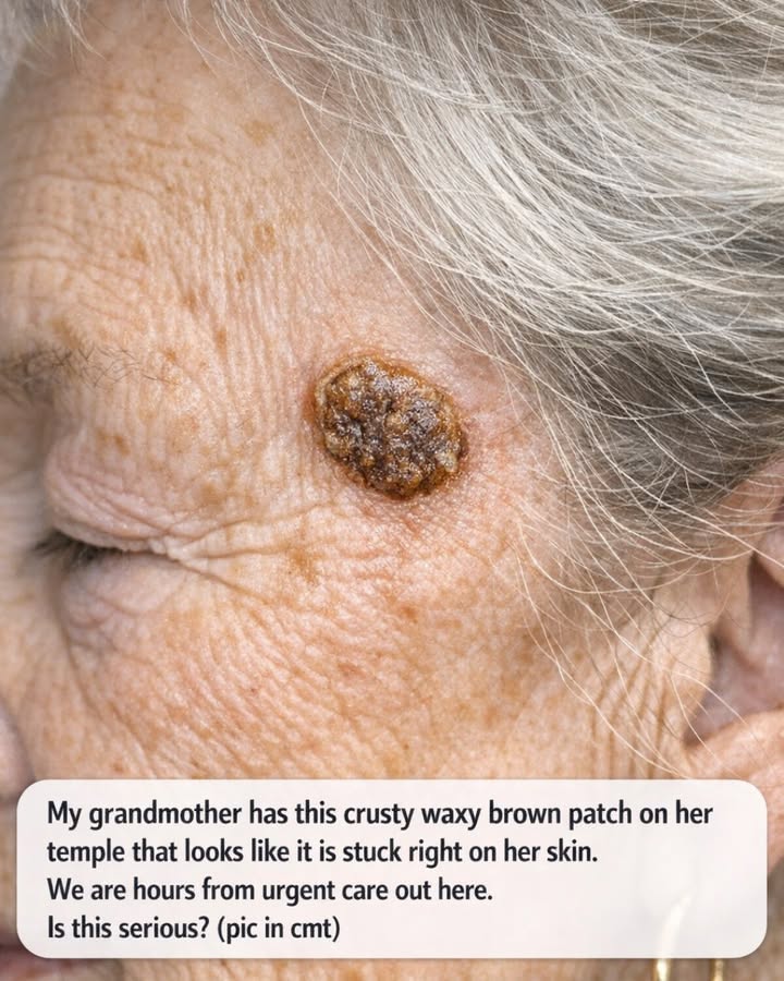
Finding a new or unusual patch on an older family member’s skin can be unsettling, especially when the spot has an unusual appearance.
A crusty, brown, waxy-looking patch on the temple may immediately make you wonder whether it is something serious. The good news is that there are several benign skin growths that can look surprisingly unusual, particularly as people get older.
One possibility is a common growth called a seborrheic keratosis. These growths often have a brown or tan color and can develop a waxy, rough, or “stuck-on” appearance. They commonly occur in middle-aged and older adults and can appear on areas including the head, neck, face, chest, and back.
However, it is important not to assume that every brown or crusty growth is a seborrheic keratosis.
A description alone cannot establish what a skin lesion is. Some skin cancers can also appear as rough, scaly, crusted, brown, or changing areas of skin. A dermatologist may need to examine the spot closely to determine what it is.
What Does a “Stuck-On” Skin Growth Mean?
The phrase “stuck on the skin” is often used to describe the appearance of seborrheic keratoses.
These growths can look as though someone placed a small piece of wax, clay, or dried material on top of the skin. They may be smooth or rough and can range in color from pale or tan to brown or very dark.
Some are relatively flat, while others become thicker and more raised over time.
They can also develop a somewhat irregular or warty surface.
This unusual appearance can understandably make family members nervous, particularly when the growth appears on the face.
But appearance alone does not tell you whether a lesion is harmless.
Why Older Adults Often Develop These Growths
Seborrheic keratoses are particularly common as people get older.
Some individuals have only one, while others develop several over time. Because these growths can become more noticeable with age, families sometimes discover them while helping an older relative with routine grooming or healthcare.
Finding one does not automatically mean that something dangerous has developed.
At the same time, a new or changing growth should not simply be dismissed because of someone’s age.
Could It Be Something Other Than a Seborrheic Keratosis?
Yes.
Several different skin conditions can produce patches, bumps, scales, or crusts.
For example, actinic keratosis can appear as a rough or scaly patch on skin that has experienced significant ultraviolet exposure. These lesions can be red, pink, gray, yellow, brown, tan, or skin-colored. Some may itch, sting, feel tender, or bleed.
Certain skin cancers can also have varied appearances.
A basal cell carcinoma may appear as a growing spot or bump, a shiny area, a brown or black growth, a waxy-looking growth, or a sore that heals and later returns. Squamous cell carcinoma may appear as a rough or scaly patch, a crusted growth, a firm bump, or a sore that does not heal.
This is why it isn’t possible to safely identify a skin lesion from a written description alone.
What Changes Should Get Extra Attention?
Certain changes make it more important to arrange a professional skin examination.
Pay particular attention if the growth:
-
Is getting noticeably larger.
-
Changes shape or color.
-
Develops several different colors.
-
Begins bleeding without an obvious injury.
-
Repeatedly forms a scab.
-
Becomes painful or tender.
-
Starts itching persistently.
-
Develops an unusual rough or scaly surface.
-
Looks significantly different from other spots on the person’s skin.
-
Becomes an open sore that does not heal or repeatedly returns.
None of these characteristics automatically means the growth is cancer.
They simply provide reasons to have it assessed rather than trying to identify it at home.
Does It Need Emergency Care?
A skin growth that has been present without major changes generally does not automatically require an emergency-room visit.
If your grandmother is otherwise well and the issue is simply an unusual-looking patch, arranging an appointment with her primary-care clinician or a dermatologist is generally more appropriate than attempting to remove it at home.
However, circumstances can change.
If the area is associated with significant bleeding that does not stop with appropriate first aid, rapidly increasing swelling, severe pain, signs of infection, or another concerning acute symptom, urgent medical evaluation may be appropriate.
For a stable skin growth without those symptoms, the more useful next step is usually professional assessment rather than panic.
Don’t Try to Peel or Cut It Off
One of the most important things to avoid is attempting to remove the growth yourself.
If the lesion is a seborrheic keratosis, a healthcare professional can remove it when necessary or when it is repeatedly irritated.
Home remedies can also make the situation harder to evaluate.
Scraping, cutting, burning, freezing, or applying strong chemicals could damage the surrounding skin and potentially change the appearance of the lesion before a doctor examines it.
That is particularly undesirable when the original diagnosis is uncertain.
What Will a Dermatologist Do?
A dermatologist will generally begin by examining the area closely.
In many cases, the appearance of a seborrheic keratosis is characteristic enough for a dermatologist to recognize it during an examination.
Sometimes, however, a growth may resemble skin cancer or another condition.
When there is uncertainty, the dermatologist may recommend removing part or all of the growth and having the tissue examined under a microscope.
This is one reason professional evaluation is more reliable than comparing photographs online.
Take a Photograph for Comparison
While waiting for an appointment, taking a clear photograph can be useful.
Try to photograph the spot in good lighting and from a consistent distance. If possible, keep a record of approximately when you first noticed it.
A photograph cannot diagnose the lesion, but it can help you notice whether the appearance changes over time.
Avoid repeatedly touching or scratching the area simply to see whether it comes off.
If the lesion becomes irritated, that can create additional changes that make it harder to assess.
What If the Spot Has Been There for Years?
A long-standing spot that has remained essentially unchanged may be less concerning than a new or rapidly changing lesion, but that does not mean you should diagnose it yourself.
Some harmless growths can remain for years, while certain skin cancers can also develop slowly.
The most useful information is the combination of history and examination.
If your grandmother remembers having the same patch for many years with little or no change, tell the clinician.
If nobody knows when it appeared, mention that as well.
Sun Exposure Is Worth Considering
The temple and other areas of the face receive regular sun exposure over a lifetime.
Long-term ultraviolet exposure is associated with several types of skin changes, including actinic keratoses and skin cancers.
That doesn’t mean a brown patch on the temple is necessarily caused by sun damage.
It simply means that facial skin deserves attention when a new or changing lesion appears.
Regular skin checks can help people notice changes earlier.
Don’t Rely on Internet Photos for a Diagnosis
Online photographs can be useful for education, but they have limitations.
Different skin conditions can look remarkably similar.
Lighting, skin tone, camera settings, image quality, and the angle of the photograph can all change how a lesion appears.
Even dermatologists sometimes need magnification or a biopsy to distinguish between similar-looking conditions.
So if a family member says, “It looks exactly like the picture I found online,” that shouldn’t be treated as a diagnosis.
Use online information to prepare questions for a healthcare professional rather than as a replacement for an examination.
A Calm and Practical Approach
If you are several hours away from urgent care, the most useful approach is to assess the situation calmly.
If your grandmother is otherwise feeling well and the patch is simply an unusual-looking growth, you can avoid disturbing it and arrange an appropriate medical appointment when possible.
If the spot is rapidly changing, repeatedly bleeding, becoming painful, or developing other concerning features, contact a healthcare professional sooner and explain the specific changes.
When calling, give useful details such as:
-
When you first noticed the spot.
-
Whether it has grown.
-
Whether its color has changed.
-
Whether it bleeds or forms a scab.
-
Whether it itches or hurts.
-
Whether it is raised or flat.
-
Whether your grandmother has had similar growths before.
-
Whether she has a history of skin cancer or significant sun exposure.
These details can help the healthcare team decide how quickly she should be evaluated.
Final Thoughts
A crusty, brown, waxy patch that appears to be “stuck” to the skin can certainly look alarming, particularly when it appears on an older person’s face.
One common possibility is a seborrheic keratosis, a generally harmless skin growth that often has a waxy, raised, or stuck-on appearance.
But that description is not enough to determine what the lesion actually is.
Other skin conditions, including actinic keratoses and skin cancers, can sometimes look similar. A lesion that grows, changes, bleeds, becomes painful, or develops a persistent rough or scaly surface deserves professional evaluation.
Until it can be examined, don’t cut, scrape, burn, freeze, or otherwise attempt to remove it at home.
If your grandmother is otherwise stable, arranging an appointment with her doctor or a dermatologist is a sensible next step. If she develops significant bleeding, severe pain, rapidly worsening swelling, signs of infection, or another acute medical problem, seek urgent medical care.
Most importantly, don’t let an alarming appearance automatically convince you that the worst has happened—and don’t let a reassuring appearance convince you that professional evaluation is unnecessary.
The safest answer comes from having the spot examined by someone qualified to identify it.